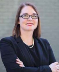
Ashley Hendrix, M.D.
Advancements in Breast Cancer Treatment
 Breast cancer is the second most common cancer in women and men worldwide. Approximately one in eight women (and 1 in 1,000 men) are diagnosed with breast cancer in their lifetime. Now for the good news: Due to medical advances in the diagnosis and treatment of breast cancer, the survival rate is dramatically improving.
As Dr. Ashley Hendrix, a board-certified breast surgical oncologist at LSU Healthcare Network and assistant professor of clinical surgery, explains, there are different elements of breast surgery, used for various reasons:
- To remove as much of the cancer as possible (breast-conserving surgery or mastectomy)
- To find out whether cancer has spread to the lymph nodes under the arm (sentinel lymph node biopsy or axillary lymph node dissection)
- To restore the breast’s shape after the cancer is removed (breast reconstruction)
- To relieve symptoms of metastatic cancer
Breast imaging technology has evolved to allow breast abnormalities and cancers to be detected before clinically evident. While this refinement enables doctors to identify problems early on, it also creates a challenge to precisely locate for surgical removal.
Once someone is diagnosed with breast cancer, tests will be done to find out the extent (stage) of the cancer. “Staging helps us to determine how severe the cancer is and how best to treat it,” Dr. Hendrx says. “The breast tumor resection is typically composed of two separate procedures. The first is to localize the tumor and the second is to remove it.” The original method was for the radiologist to place guide wire at the center of the tumor to navigate the surgeon to the area of resection. “Wire localization required that the wire is placed near the mass immediately prior to surgery,” she adds. “The patient was then transported to the operating room with the wire protruding from their breast. This caused complications in scheduling as well as limited mobility and discomfort for some patients.”
Radioactive Seed Localization offers an alternative to the wire and a unique set of benefits. In the RSL procedure, a radiologist places a radioactive seed measuring 5 mm (about the size of a grain of rice) near the abnormal mass one to five days prior to surgery. Composed of a minimal amount of radioactive iodine, the seed is encased in a titanium shell to prevent the risk of exposure,” Dr. Hendrx says. “Although RSL offers a more accurate, comfortable and convenient alternative to the traditional method, there are drawbacks. Radioactive seeds come with protocols and regulations. Although an improvement over the wire, RSL still requires a lot of tracking and has time restraints when it comes to scheduling surgery.”
New Tools for Better Targeting
Sentimag, a device that uses alternating magnetic waves to identify objects, was approved by the FDA in 2015. Used in conjunction with the Magseed, a non-radioactive, iron oxide seed that can be placed up to 30 days in advance of surgery, this reduces times constraints and difficultly in navigating the difficulty of surgical scheduling.
The Sentimag system greatly enhances convenience for both patients and clinicians. “This is a great advance for breast cancer treatment,” Dr. Hendrix says. “With Magseed, we can accurately remove the breast tumor, leaving behind as much healthy tissue as possible and make the experience as easy as possible for patients.”
There are other alternatives used elsewhere that are non-radioactive, such as the SAVI SCOUT and Faxitron localizer.
More Advancements
Dr. Hendrix works with a team of plastic and reconstructive surgeons and says that oncoplastic surgery and nipple sparing mastectomies have significantly improved cosmetic results. “Many patients undergo chemotherapy prior to surgery, allowing for possible down-staging of cancer,” she says. “If down-staging does not occur or if the patient prefers mastectomy, there are several different types of mastectomies in which nipple preservation may be a possibility.”
At LSU Healthcare Network, Dr. Hendrix works with colleagues who specialize in pre-flap reconstruction, including DIEP flap breast reconstruction (Deep Inferior Epigastric Perforators), one of the most advanced forms of breast reconstructive surgery available today. It is a microsurgical technique that requires a highly skilled surgeon. The procedure uses a flap of excess fat, skin and blood vessels from the patient’s lower abdomen to construct a new breast made of soft, warm, living tissue, while leaving the abdominal muscles intact. “Because reconstructive has been preforming this type of reconstructive surgery since 1994, we do more tissue reconstruction than the rest of the United States,” she says.
Dr. Hendrix joined LSU Healthcare Network after spending three years in private practice in Tennessee after completing her Fellowship in breast surgery. She says when she came to LSU it felt like she was coming home and appreciated the support of her colleagues and faculty, all of whom she feels are focused on patient care. She began her career in medicine as a biomedical engineer but decided she missed interacting with patients. While in medical school she had the opportunity to shadow Dr. Elizabeth Pritchard at the University of Tennessee Health Science Center and that changed her life. “I got a close-up look at patient care; got to meet with survivors, and got to know the patients,” Dr. Hendrix says. “It was then that I decided I wanted to be just like Dr. Pritchard, who has remained my role model and mentor.” She recently expanded her practice to the LSU Healthcare Network Westbank location (1111 Medical Center Blvd., Ste. S640, Marrero).
Ashley Hendrix, M.D.
LSU Healthcare Network
1111 Medical Center Blvd., Suite S640
Marrero, LA 70072
(504) 412-1390
MEDICAL SCHOOL: The University of Tennessee College of Medicine
RESIDENCY: University of Tennessee
FELLOWSHIP: University of Tennessee Southwestern Medical Center
CERTIFICATIONS: Board Certified General Surgery, Hidden Scar Surgery
FELLOWSHIP: Breast Surgery
